UT Health Northeast now offers technology to better detect breast cancer in women with dense tissue
Published 4:16 pm Friday, August 19, 2016
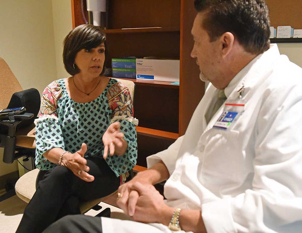
- Patient Kelly Nichols of Bullard and Dr. Don Wells talk about family medical history before an Invenia Automated Breast Ultrasound System screening Thursday August 18, 2016 at UT Health Northeast in Tyler. The new technology is used to increase early detection of breast cancer and is especially helpful for women with dense breast tissue. (Sarah A. Miller/Tyler Morning Telegraph)
COSHANDRA DILLARD, cdillard@tylerpaper.com
Kelly Nichols, 49, of Bullard, has been getting mammograms since she was 19 because she felt a knot in a breast. But Thursday was the first time to experience a screening that better detects breast cancer in patients like her who have dense breast tissue.
In the last three or four years, doctors have recognized that the mother of two has dense breast tissue, which is difficult to detect with a mammogram, so she has been getting a sonogram in addition to a mammogram to increase the chances of spotting cancer.
Now, Mrs. Nichols is able to get more thorough detection locally using a new 3D system at UT Health Northeast known as ABUS (Automated Breast Ultrasound System).
Although the technology has been around for a few years, UT Health’s system is the first for East Texas, making it possible for local women like Mrs. Nichols to better identify cancer that may be lurking amidst other tissue.
In recent years, studies published in the American Journal of Roentgenology, found the 3D technology could detect up to 35.6 percent more cancers compared to mammograms alone.
Radiologist Dr. Don Wells said about one-third of dense breast tissue can’t be seen with mammography only. In imaging, the fibroglandular tissue is white, just as cancerous lumps.Radiologist Dr. Don Wells said dense breast tissue is common in women.
Wells said dense breast tissue is common in women. He said 40 percent of women younger than 50 and 30 percent of women older than 50 have dense breast tissue. It could change over time.
“As women get older, their breast tissue becomes fattier,” Wells said. “Women with dense breast tissue are likely to have dense breast tissue for the rest of her life. It will become less dense in most women over time, but it’s also influenced by a woman who’s on hormonal replacement therapy or women who drink a lot of caffeine. There are a lot of factors involved, including genetics.”
Henda’s Law, passed by Texas legislators in 2011, requires doctors to notify patients if they have dense breast tissue. It’s then up to the patient to decide if she’ll have a supplemental screening.
For Mrs. Nichols, being proactive about screenings is critical, as her mother was diagnosed with breast cancer five years ago. But despite serious concern, she content when she showed up at UT Health Northeast’s Breast Center Thursday for the procedure.
“This is good. I’m not nervous,” Mrs. Nichols said before the procedure, which lasted about 30 minutes.
Because of Mrs. Nichols’ history, doctors had previously recommended she get an MRI. MRIs are sensitive to detecting cancers, but the modality has a lower specificity, meaning it may be more difficult to differentiate benign lesions from malignant lesions.
The cost of the 3D technology varies based on insurance plans, but is notably less expensive than an MRI.
When Mrs. Nichols learned from a friend that UT Health Northeast was offering the 3D ultrasound to improve chances of catching small and invasive cancer, she was eager to give it a try.
“You can’t put stuff off like this,” she said. “I wanted to do what’s best for me and my family, so if the doctor wanted me to do it, I’d do it, regardless of the price … I don’t want to not take care of it. It’s when you put it off that something happens … Not knowing is more scary than knowing something’s there.”
Wells said the procedure is appropriate for women with dense breast tissue who have had negative mammograms, and generally, women who have not had prior procedures.
Women may be more comfortable during the procedure, since it uses only mild compression when the imaging device is placed on the breast.
Mrs. Nichols said her mother has recovered well from breast cancer, thanks to early detection. And with a 22-year-old daughter, it’s important for her to lead by example.
“She knows this is going on with me,” she said. “If it gets recommended that something be done, then we will get it done.”


-
eEdition
-
-
